Pet Radiology Services in Ridgefield, CT

Get clear answers about your pet’s health with advanced radiology services from Ridgefield Veterinary Center. Since 1955, we’ve provided expert diagnostic imaging for dogs and cats throughout Ridgefield, Redding, Wilton, Danbury, South Salem, and North Salem. Our digital x-ray technology delivers immediate, high-quality images during thirty-minute appointments. For complex cases, we consult with Dr. Victor Rendano, a board-certified veterinary radiologist, ensuring expert interpretation. Our Fear Free certified team makes imaging as comfortable as possible for your pet, using gentle techniques that minimize stress while obtaining the diagnostic information needed for accurate treatment.
Why Pet Radiology Services Matter for Accurate Diagnosis
X-rays reveal what physical examination cannot detect, providing essential information about bones, organs, and internal structures. Radiology is fundamental to accurate veterinary diagnosis and effective treatment planning.
Pet radiology services diagnose fractures and bone injuries, detect tumors and masses, evaluate heart and lung disease, identify foreign objects in the stomach or intestines, and assess arthritis and joint problems.
Immediate Visual Diagnosis – Digital x-rays show bone fractures, joint abnormalities, bladder stones, enlarged organs, lung infections, heart enlargement, spinal problems, and foreign objects your pet may have swallowed.
Surgical Planning – Before orthopedic surgery, x-rays show the exact location and severity of fractures or joint damage, allowing precise surgical planning and better outcomes.
Emergency Care – When pets are injured or suddenly ill, x-rays quickly identify life-threatening conditions like internal bleeding, organ rupture, or intestinal obstructions requiring immediate surgery.
Chronic Disease Management – For pets with heart disease, arthritis, or cancer, serial X-rays monitor disease progression and treatment effectiveness over time.
Pre-Anesthetic Screening – Chest x-rays before surgery evaluate heart and lung health, identifying conditions that could cause anesthetic complications.
Board-Certified Expertise – Complex cases benefit from consultation with Dr. Victor Rendano, ensuring even challenging X-rays receive expert interpretation for accurate diagnosis.
What to Expect During Your Pet’s Radiology Appointment
Pet radiology at Ridgefield Veterinary Center provides high-quality diagnostic imaging during efficient thirty-minute appointments, with immediate results guiding treatment decisions.
Before the X-Ray (5 minutes)
We discuss your pet’s symptoms and why x-rays are recommended. Our veterinarians explain which body areas will be imaged and what we’re looking for. For anxious pets, our Fear Free trained staff uses calming techniques. Most pets tolerate X-rays well with gentle restraint, but sedation is available if needed for patient comfort or image quality.
During the X-Ray (10-15 minutes)
- Positioning: We carefully position your pet to obtain clear images of the affected area. Multiple views are typically needed for complete evaluation. Our experienced staff works quickly and gently to minimize restraint time.
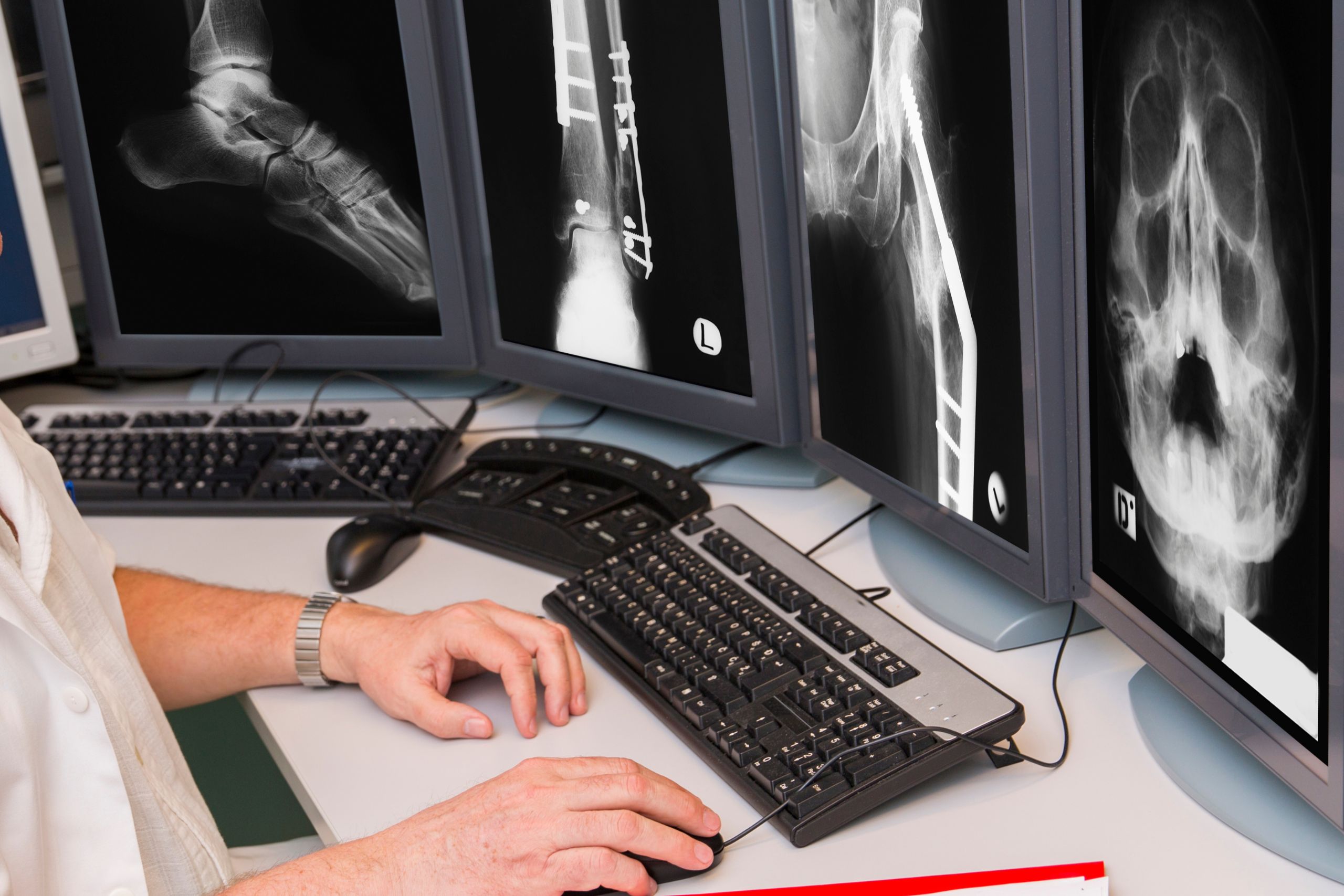
- Image Acquisition: Our digital X-ray system captures high-quality images within seconds. Unlike traditional film x-rays that required chemical processing, digital images appear immediately on computer screens. We can adjust brightness, contrast, and magnification to enhance the visualization of subtle abnormalities.
- Multiple Views: Most examinations require 2-4 x-rays from different angles. Orthopedic cases may need both affected and normal limbs for comparison. Chest x-rays typically include two views to thoroughly evaluate the heart and lungs.
After the X-Ray (10-15 minutes)
Our veterinarians review images immediately and discuss findings with you. We explain what x-rays show in understandable terms, recommend treatment based on radiographic findings, discuss prognosis and expected outcomes, and determine if additional imaging or testing is needed.
For complex cases, we consult with Dr. Rendano, board-certified veterinary radiologist, for expert interpretation. Your pet can go home immediately unless sedation was used.
Total appointment time: Thirty minutes provides complete radiology services efficiently, with immediate results guiding treatment decisions.
When Should Your Pet Receive Radiology Services?
X-rays are essential diagnostic tools for many conditions affecting dogs and cats. Understanding when radiology is needed ensures your pet receives timely, appropriate care.
Common Conditions Requiring X-Rays
| Condition | What X-Rays Reveal | Treatment Guidance |
|---|---|---|
| Limping/Lameness | Fractures, arthritis, bone tumors, joint problems | Determines if surgery, medication, or rest is needed |
| Difficulty Breathing | Heart enlargement, lung disease, fluid accumulation, tumors | Guides cardiac or respiratory treatment |
| Vomiting/Diarrhea | Foreign objects, intestinal obstruction, organ enlargement | Identifies if emergency surgery is required |
| Trauma/Injury | Broken bones, internal injuries, organ damage | Assesses injury severity and surgical needs |
| Coughing | Heart disease, tracheal collapse, lung infections | Determines if heart or lung medication is needed |
| Abdominal Pain | Bladder stones, organ enlargement, masses, obstructions | Identifies the cause of pain requiring treatment |
| Urinary Problems | Bladder stones, enlarged prostate, kidney disease | Shows stones requiring surgical removal |
| Weight Loss | Organ abnormalities, masses, heart disease | Identifies underlying causes |
| Chronic Arthritis | Joint degeneration, bone spurs, hip dysplasia | Guides pain management and treatment |
| Pre-Surgical Screening | Heart and lung health, overall body condition | Ensures safe anesthesia |
Signs Your Pet Needs X-Rays Now
- Limping, lameness, or inability to bear weight on a leg
- Difficulty breathing or rapid, labored breathing
- Persistent vomiting, especially after eating
- Abdominal swelling or signs of pain
- Severe lethargy after trauma or injury
- Coughing that doesn’t resolve
- Straining to urinate or blood in urine
- Sudden paralysis or inability to walk
- Suspected ingestion of foreign objects
- Falls or vehicle accidents
Routine Radiology for Senior Pets
Senior pets benefit from screening x-rays during wellness exams:
Heart and Lung Evaluation:Chest x-rays detect early heart enlargement or lung disease before symptoms appear, allowing proactive treatment.
Arthritis Assessment: Skeletal x-rays identify arthritis severity, guiding pain management and mobility support.
Cancer Screening: X-rays can detect bone tumors or masses requiring further evaluation.
Pre-Surgical Radiology Requirements
X-rays are often required before surgical procedures:
Orthopedic Surgery: X-rays show fracture location, joint damage, or bone abnormalities requiring surgical correction.
Dental Procedures: Dental x-rays evaluate tooth roots and jawbone health before extractions or oral surgery (when available).
Pre-Anesthetic Screening: Chest x-rays ensure the heart and lungs are healthy enough for anesthesia.
How Ridgefield Veterinary Center Provides Pet Radiology Differently
Advanced Digital Radiology Technology
Our modern digital X-ray system provides immediate high-quality images viewable within seconds, enhanced image manipulation for better diagnostic visualization, lower radiation exposure ensuring patient safety, permanent digital storage in medical records, and easy sharing with specialists for consultation. Digital imaging surpasses traditional film X-rays in speed, quality, and diagnostic capability.
Board-Certified Radiologist Consultation
Dr. Victor Rendano, board-certified veterinary radiologist, provides expert consultation for complex cases. Through Veterinary Multi-Imaging (VMI), we access decades of specialized radiology expertise, ensuring accurate interpretation even in challenging diagnostic situations. Dr. Rendano has published over 100 papers, lectured internationally, and provides radiograph interpretation, ultrasonography consultation, and advanced imaging guidance.
Fear Free Imaging Techniques
Our Fear Free certification ensures radiology procedures minimize stress through gentle restraint without force, calming pheromones in the radiology suite, treats and positive reinforcement during positioning, skilled technicians working quickly and efficiently, and sedation available when needed for anxious or painful pets. Calm pets produce better quality images with less motion artifact.
Comprehensive Radiology Services
We provide complete imaging for dogs and cats, including orthopedic x-rays for limbs, joints, and spine, chest x-rays evaluating heart and lungs, abdominal x-rays assessing organs and detecting foreign objects, skull x-rays for dental and neurological evaluation, and full-body surveys when needed for trauma or cancer screening.
Immediate Results and Treatment Planning
Digital technology means no waiting for film processing. Our veterinarians review x-rays with you during the appointment, explain findings clearly, recommend treatment based on radiographic diagnosis, and often begin therapy immediately. Fast diagnosis means faster treatment and better outcomes.
70 Years of Imaging Experience
Since 1955, our expertise in veterinary radiology has meant skilled positioning for optimal images, recognition of subtle abnormalities, knowledge of breed-specific variations, and integration with comprehensive medical care.
Comprehensive Veterinary Services Utilizing Radiology
Pet radiology integrates with multiple veterinary services, supporting complete diagnostic care:
Pet Wellness Exams in Ridgefield, CT – Wellness exams for senior pets may include screening, x-rays, and detection of heart disease, arthritis, or tumors before symptoms appear.
Pet Surgery Services in Ridgefield, CT – Pre-surgical x-rays guide orthopedic procedures. Post-operative imaging confirms proper healing and hardware placement.
Pet Diagnostic Services in Ridgefield, CT – Radiology combines with blood work and other laboratory testing for a comprehensive diagnosis of complex conditions.
Senior Pet Care in Ridgefield, CT – Older pets benefit from chest and skeletal x-rays, monitoring age-related diseases, and guiding geriatric care.
Fear Free Veterinary Care – Our certification ensures all radiology procedures use gentle techniques, creating positive experiences for anxious pets.
Schedule Your Pet’s Radiology Appointment Today
Get accurate diagnostic answers with expert pet radiology from Ridgefield Veterinary Center. Our digital x-ray technology and board-certified radiologist consultation ensure precise diagnosis in efficient thirty-minute appointments.
Three Easy Ways to Schedule:
📞 Call us directly: 203-438-2658 – Our team discusses diagnostic needs and schedules your appointment.
🖥️ Book online now – Request your diagnostic appointment 24/7 through our convenient scheduling system.
📍 Visit us: 722 Danbury Road, Ridgefield, CT 06877 – Monday-Friday 8:00 AM – 5:00 PM (Closed 1-2 PM daily)